
EKG Lead Placement
Where every ECG electrode goes: the 12-lead, the 5-lead and 3-lead monitor, and the right-sided and posterior leads, from AHA statements, guidelines and peer-reviewed papers. Choose an electrode to see its landmark.
Chest electrodes are placed by bony landmarks, limb electrodes on the arms and legs. Choose one here or in the list.
12-lead EKG placement
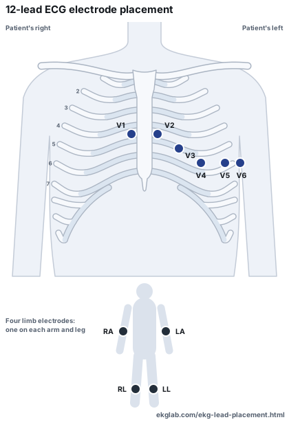
A standard ECG uses ten electrodes, six on the chest1 and four on the limbs.14 The right leg electrode is a reference, so nine of the ten form the twelve leads. These are the positions in the AHA/ACCF/HRS recording standard, which this page calls the AHA standard.
- Fourth intercostal space, right sternal border1IEC label C1
- Fourth intercostal space, left sternal border1IEC label C2
- Midway between V2 and V41IEC label C3
- Fifth intercostal space, midclavicular line1IEC label C4
- Right arm, below the shoulder1IEC label R
- Left arm, below the shoulder1IEC label L
- Left leg, below the hip1IEC label F
The limb wording is the AHA's 1975 statement, which the standard reports,1 adding that placement along a limb can alter the ECG.1 The IEC labels are from Paul and Jacob and the SCST guideline.115
How to find V1, then the rest in order
The AHA standard names the landmarks and warns that electrodes positioned without reference to the bones often end up in a vertical line.11 The SCST guideline, from the UK's professional body, describes how to reach the landmarks. This is its method.
- A finger is run down the sternum5 until it meets a bony ridge,5 the angle of Louis. The second costal cartilage joins the sternum there.18
- Sliding down and to the patient's right finds the second intercostal space.5 From there, count down to the third and the fourth.5
- In the fourth space, slide toward the sternum until its edge is felt. V1 goes there.5
- Repeat on the patient's left for V2. The guideline says to count again rather than place V2 beside V1, because the two sides may be offset.5
- V4 goes in the fifth intercostal space, in line with the midpoint of the clavicle.5
- V3 goes midway between V2 and V4,5 which is why V4 is found first.
- V5 and V6 go in horizontal alignment with V4.5 The AHA standard discourages following the fifth space around the chest instead.1
Anatomy varies. In a study of 85 adult sterna, the sternal angle did not match the second costal notch in one.1919
Position and skin
The AHA standard describes the recording as historically made with the patient supine, generally with a pillow under the head,1 and says tracings recorded sitting or upright should not be considered equivalent to standard supine ECGs.1 It also says skin preparation by cleaning and gentle abrasion can reduce noise.1 The monitoring standards describe cleansing the skin, wiping it with a dry cloth and clipping excess hair.44
Breast tissue
The AHA standard recommends electrodes stay under the breast in women until more studies are available.1 The SCST guideline calls this the convention for V4, V5 and V6.5 Rautaharju 1998 recommended the opposite.17
Serial tracings
The AHA standard says serial tracings in acute or subacute care settings should use some form of skin marking when electrodes cannot stay on.1
Try it: place V1 to V6
Click where V1 goes
Count the ribs. They are numbered on the patient's right.
Answer from a list instead of the picture
The 12 ECG leads, and where they come from
An electrode is the sensor on the skin. A lead is a view worked out from the electrodes, which is why ten electrodes give twelve leads. The limb leads lie in the frontal plane,1 and V1 to V6 progress leftward and slightly inferiorly across the chest.1
| Limb leads, from the arm and leg electrodes | |
|---|---|
| Lead I | Left arm compared with right arm1 |
| Lead II | Left leg compared with right arm1 |
| Lead III | Left leg compared with left arm1 |
| aVR, aVL, aVF | RA, LA and LL, each compared with the average of the other two.1 See the augmented limb leads. |
| Precordial leads, from the chest electrodes | |
|---|---|
| V1 to V6 | Each chest electrode compared with Wilson's central terminal,1 the average of RA, LA and LL.1 See the 12-lead ECG. |
Common EKG lead placement mistakes
The AHA monitoring standards report that in studies of hospital monitoring, electrode misplacement ranged between 20% and 80%.4 When 30 experienced technicians were measured, V1 and V2 were displaced upward by more than 0.625 inch in more than half of applications.15 When 120 staff marked diagrams, V1 and V2 often landed in the second space.16 The AHA standard says chest electrode variation of as little as 2 cm can cause important diagnostic errors,1 particularly in statements about anteroseptal infarction and hypertrophy.1
| Mistake | What the sources say it can show |
|---|---|
| V1 and V2 too high | A common error, in the second or third space:1 poor R-wave progression or false signs of anterior infarction.1 |
| V5 and V6 too low | Also common, in the sixth space or lower:1 changed amplitudes, which are used in diagnosing hypertrophy.1 |
| Left and right arm wires swapped | Lead I inverted, leads II and III switched, aVR and aVL switched, aVF unchanged, no change in the chest leads.11 Lead I and V6 stop matching.1 |
| Right arm and right leg wires swapped | Very low amplitude in lead II alone.1 The chest leads change too, because the neutral wire has moved.10 With the leg electrodes on the torso that flat lead II may not appear.11 |
| Left arm and left leg wires swapped | Harder to spot: often a subtle shift in axis, and lead III inverted.1 |
| Left arm and right leg wires swapped | Lead III appears as a near flat line.11 Moving the right leg wire to an arm also changes the chest leads.10 |
| Left leg and right leg wires swapped | No significant change, because the voltages in the two legs are almost identical.11 |
| Each arm wire swapped with a leg wire | Lead I appears as a near flat line, and all twelve leads are affected.11 |
| Chest wires swapped | V1 with V2, or V2 with V3: a reversal of R-wave progression that can look like anteroseptal infarction.1 |
The AHA statement list has its own technical statements for these: extremity electrode reversal and misplaced precordial electrodes.22 The standard also reports that chest misplacement can change computer statements in up to 6% of recordings.1
5-lead EKG placement
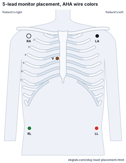
A 5-lead monitor uses five electrodes and gives seven leads: I, II, III, aVR, aVL, aVF and V.84 The limb electrodes go on the torso to allow patient movement while reducing artifact.4 Only one chest lead is possible at a time.9
- Right infraclavicular fossa, medial to the deltoid1White (AHA), red (IEC)
- Left infraclavicular fossa, medial to the deltoid1Black (AHA), yellow (IEC)
- Left anterior axillary line, midway between the costal margin and iliac crest1Red (AHA), green (IEC)
These are the Mason-Likar positions as the AHA standard describes them, the placement a published ICU monitoring study used.8 The standard adds that more recent applications place the arm electrodes over the outer clavicles.1
3-lead EKG placement
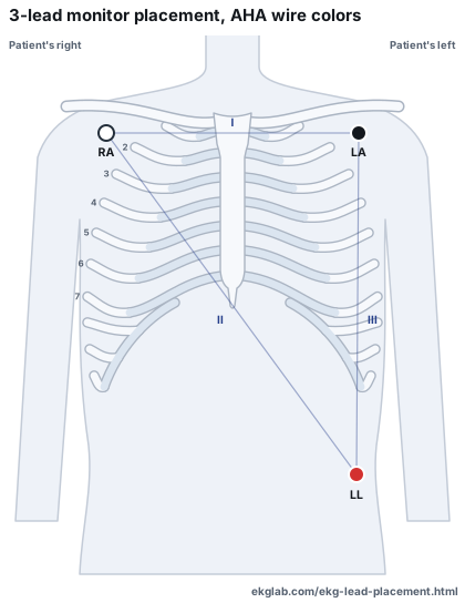
A 3-lead monitor uses two electrodes at a time and the third as a ground,9 so it shows lead I, II or III, one at a time.9 The electrodes usually go on the torso near the limb they stand for.9
- Right infraclavicular fossa, medial to the deltoid1White (AHA), red (IEC)
- Left infraclavicular fossa, medial to the deltoid1Black (AHA), yellow (IEC)
- Left anterior axillary line, midway between the costal margin and iliac crest1Red (AHA), green (IEC)
Francis 2016 says only that the electrodes go near the corresponding limb, so the sites drawn are the 5-lead's. The 3-electrode facts on this page rest on that one review, the only source read that sets them out.
Which lead is monitored
The AHA monitoring standards say that for arrhythmia monitoring V1 is commonly selected, for its help in telling VT from aberrancy,4 and that a precordial lead, V1 or V6, is needed to assess ventricular arrhythmias.4 In children, lead II is commonly the primary lead.4
Torso electrodes and the diagnostic 12-lead
The AHA standard says rhythm diagnosis is not adversely affected by monitoring lead placement.1 Other things change: electrodes on the trunk do not provide standard limb leads,1 the augmented and chest leads are altered too,1 and the differences can include false infarction criteria in both directions.1 So the standard says such a tracing cannot be considered equivalent to a standard ECG for all purposes, should not be used for serial comparison with one, and must be clearly labeled.111 The SCST guideline says limb electrodes must not be placed on the torso for a standard recording.5 The monitoring standards list moving the limb wires from the torso to the limbs as a skill for recording a standard 12-lead from a monitor.4
Right-sided EKG leads
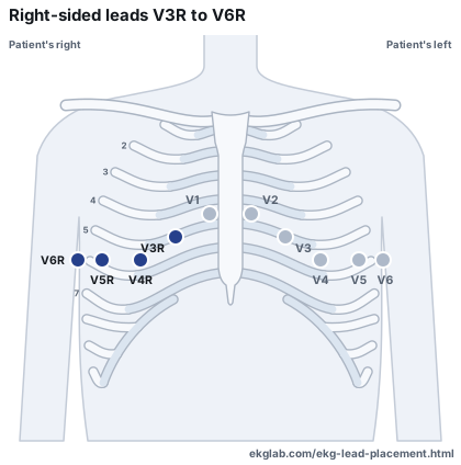
V3R to V6R go on the right of the chest, in mirror image to the standard positions.1 Standard V1 can be considered V2R, and standard V2 can be considered V1R.11 The AHA standard treats them as additional leads, not routine ones.1
- Midway between V1 and V4R12
- Fifth intercostal space, right midclavicular line12
- Level with V4R, right anterior axillary line1
- Level with V4R, right midaxillary line13
A recording from moved electrodes needs labeling. The SCST guideline says any non-standard position must be labelled on the recording,5 and AHA Part VI says machines should be able to label V3R and V4R.32
Posterior EKG lead placement
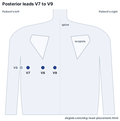
V7, V8 and V9 go on the back of the left chest. The AHA standard describes them as proposed additional leads,1 each in the same horizontal plane as V6,1 and not for routine recording.1
- Left posterior axillary line1IEC label C7
Without a device that records them, the SCST guideline moves V4, V5 and V6 to V7, V8 and V9 for a second recording, clearly annotated.55 The AHA statement list includes posterior electrodes as a technical statement.2
15-lead and 18-lead ECGs
The names are not fixed. One study's 15-lead added V4R, V8 and V9,20 a pediatric 15-lead adds V3R, V4R and V7,12 and an 18-lead has been described as the 12 plus V3R to V5R and V7 to V9.21
EKG wire colors and labels
The AHA standard says color coding is a feature of manufacturing standards.1 Two codes are in use. This table is from Paul and Jacob, a peer-reviewed review.11 The SCST guideline gives the same IEC colors55 and notes that colors may vary by manufacturer.5
| Electrode | AHA label | AHA color | IEC label | IEC color |
|---|---|---|---|---|
| Right arm | RA | White | R | Red |
| Left arm | LA | Black | L | Yellow |
| Right leg | RL | Green | N | Black |
| Left leg | LL | Red | F | Green |
| Chest | V1 to V6 | Brown, then red, yellow, green, blue, orange and purple for V1 to V6 | C1 to C6 | White, then red, yellow, green, brown, black and purple for C1 to C6 |
Where the standards differ
The sources do not agree on everything. Where they differ, this page shows each one rather than picking.
| Point | One source | Another |
|---|---|---|
| Patient position | AHA standard: supine, with sitting or upright tracings not equivalent.1 | SCST: about 45 degrees.5 |
| Limb electrodes | AHA standard, reporting the AHA's 1975 statement: on the arms and legs below the shoulders and hips, not necessarily at the wrists and ankles.11 | SCST: near the wrists and ankles.5 |
| Alcohol on the skin | AHA monitoring standards, for monitoring electrodes: should not be used.4 | SCST, for a 12-lead recording: lists an alcohol-based wipe as one way to cleanse.5 |
| Level of V7 to V9 | Fourth Universal Definition (2018): the fifth intercostal space.6 The Fifth (2026) gives no level.7 | AHA standard: the plane of V6.1 SCST: the level of V4.5 Birnbaum 2020 points out the difference.14 |
| Electrodes and breast tissue | AHA standard and SCST: under the breast.1 | Rautaharju 1998: on the breast.17 |
Where these positions come from
- EinthovenDefined the frontal plane limb leads.1
- 1934Wilson and colleagues devised a central terminal from the limb electrodes as a new reference.1
- 1938A joint committee of the AHA and the Cardiac Society of Great Britain and Ireland first standardized the chest positions.1
- 1942Goldberger produced the augmented limb leads.1
- 1966Mason and Likar's torso position, used to reduce motion noise in exercise and ambulatory recording.1
- 1975An AHA statement recommended limb electrodes on the arms and legs, not necessarily at the wrists and ankles.1
- 2007The AHA, ACCF and HRS standard, the source for this page's 12-lead positions.
- 2017The AHA monitoring standards, updating those of 2004.
- 2024The current SCST guideline, which follows the AHA positions.5
EKG lead placement charts
Every setup on this page as a chart. Use them free for study and teaching, with a link back to this page.
Now read what those leads record
Practice on real 12-lead ECGs and rhythm strips, with calipers and a magnifier. Pro adds the 12-Lead and STEMI Workups, Telemetry Watch on a live bedside monitor, True Sim, the rhythm simulator built only from real patient recordings, and ARIA, which marks P, QRS, ST and T on any beat you click.
EKG and ECG lead placement questions
Where do the leads go on a 12-lead EKG?
Ten electrodes go on the body. In the AHA standard, V1 and V2 sit in the fourth intercostal space at the right and left sternal borders, V4 in the fifth intercostal space on the midclavicular line, V3 midway between V2 and V4, and V5 and V6 level with V4 on the anterior axillary and midaxillary lines. The four limb electrodes go on the arms and legs.
How many electrodes does a 12-lead ECG use?
Ten: six on the chest and four on the limbs. The right leg electrode is an electronic reference, so nine of the ten form the twelve leads.
What is the difference between an electrode and a lead?
An electrode is the sensor on the skin. A lead is a view worked out from the electrodes. The word is often used for the sticker and its wire too, which is where the confusion starts.
Where does V1 go?
In the fourth intercostal space at the right sternal border. The SCST guideline finds it by feeling for the angle of Louis, the ridge on the sternum, then counting down from the second intercostal space to the fourth.
Where do the leads go on a 5-lead monitor?
Four electrodes go on the torso near their limbs. In the Mason-Likar position the arm electrodes are in the infraclavicular fossae and the left leg electrode is midway between the costal margin and iliac crest in the anterior axillary line. The right leg electrode acts as the ground, and the fifth electrode goes on a chest position, usually V1.
How does a 3-lead monitor work?
It uses two electrodes at a time and the third as the ground, usually with the electrodes on the torso near their limbs. It shows lead I, II or III, one at a time.
Do all ECG wires use the same colors?
No. In the AHA code RA is white, LA black, RL green and LL red. In the IEC code R is red, L yellow, N black and F green. Colors can also vary by manufacturer.
Can limb electrodes go on the torso for a 12-lead ECG?
Monitors place them there. The AHA standard says a tracing recorded that way cannot be considered equivalent to a standard ECG for all purposes and must be clearly labeled. The SCST guideline says limb electrodes must not be placed on the torso for a standard recording.
Is this page clinical advice?
No. It is a study guide that reports where published standards place the electrodes. Follow your own institution's procedure and your monitor's instructions.
Sources
Point at any number on this page, or tap it, to read the source's own words and the page they are on. No manufacturer's material or general website is used.
Standards and guidelines
- Kligfield P, Gettes LS, Bailey JJ, et al. Recommendations for the standardization and interpretation of the electrocardiogram: part I: the electrocardiogram and its technology. Circulation. 2007;115(10):1306-1324. Read the sourceThe AHA, ACCF and HRS standard for recording an ECG. This page calls it the AHA standard. It is the source for the 12-lead positions.
- Mason JW, Hancock EW, Gettes LS, et al. Recommendations for the standardization and interpretation of the electrocardiogram: part II: electrocardiography diagnostic statement list. Circulation. 2007;115(10):1325-1332. Read the sourceThe standard list of ECG statements, which includes the technical statements for moved and reversed electrodes.
- Wagner GS, Macfarlane P, Wellens H, et al. AHA/ACCF/HRS recommendations for the standardization and interpretation of the electrocardiogram: part VI: acute ischemia/infarction. Circulation. 2009;119(10):e262-e270. Read the sourceThe standard for ischemia and infarction. Used here only for labeling right-sided leads.
- Sandau KE, Funk M, Auerbach A, et al. Update to practice standards for electrocardiographic monitoring in hospital settings: a scientific statement from the American Heart Association. Circulation. 2017;136(19):e273-e344. Read the sourceThe AHA statement on bedside and telemetry monitoring, updating the 2004 practice standards.
- Campbell B, Richley D, Ross C, Eggett CJ. Clinical guidelines by consensus: recording a standard 12-lead electrocardiogram. An approved method by the Professional Body for Cardiac Scientists (SCST). Document CS3, version 4.1. September 2024. Read the sourceThe UK professional body's recording guideline, which follows the AHA positions and describes how to find them.
- Thygesen K, Alpert JS, Jaffe AS, et al. Fourth universal definition of myocardial infarction (2018). Eur Heart J. 2019;40(3):237-269. Read the sourceThe ESC, ACC, AHA and WHF definition. Used here only for how it words the posterior leads.
- Mills NL, Newby LK, Zaman S, et al; ESC/ACC/AHA/WHF Universal Definition Document Advisory Group. Fifth universal definition of myocardial infarction (2026). Glob Heart. 2026;21(1):64. Read the sourceThe current definition. Used here only to show it names V7 to V9 without giving a level.
Peer-reviewed papers
- Drew BJ, Harris P, Zègre-Hemsey JK, et al. Insights into the problem of alarm fatigue with physiologic monitor devices: a comprehensive observational study of consecutive intensive care unit patients. PLoS One. 2014;9(10):e110274. Read the sourceA study of ICU monitoring that states its electrode configuration.
- Francis J. ECG monitoring leads and special leads. Indian Pacing Electrophysiol J. 2016;16(3):92-95. Read the sourceA review of monitoring lead systems. The only source read here that sets out the 3-electrode system.
- Batchvarov VN, Malik M, Camm AJ. Incorrect electrode cable connection during electrocardiographic recording. Europace. 2007;9(11):1081-1090. Read the sourceA review of electrode cable reversals. Abstract read, not the full text.
- Paul A, Jacob J. Electrocardiographic lead reversals. Indian Pacing Electrophysiol J. 2023;23(6):205-213. Read the sourceA review of lead reversals. Its Table 1 gives the IEC and AHA labels and colors.
- Mayourian J, Ghelani S, Triedman JK. Artificial intelligence-enabled electrocardiograms: do 15-lead studies improve model performance? JACC Adv. 2025;4(7):101866. Read the sourceStates the positions of V3R, V4R and V7 as used for a 15-lead ECG in pediatric cardiology.
- Morales-Arteaga JL, Meghdadi A, Wu D, et al. Alternative lead ECG placements. Ann Noninvasive Electrocardiol. 2026;31(3):e70176. Read the sourceA review of lead placements other than the standard. Used for V6R.
- Birnbaum Y, Fiol M, Nikus K, et al. A counterpoint paper: comments on the electrocardiographic part of the 2018 Fourth Universal Definition of Myocardial Infarction. Ann Noninvasive Electrocardiol. 2020;25(5):e12786. Read the sourceEndorsed by two electrocardiology societies. It sets out the disagreement over the level of V7 to V9.
- Wenger W, Kligfield P. Variability of precordial electrode placement during routine electrocardiography. J Electrocardiol. 1996;29(3):179-184. Read the sourceMeasured where 30 experienced technicians placed chest electrodes against marked sites. Abstract read, not the full text.
- Rajaganeshan R, Ludlam CL, Francis DP, Parasramka SV, Sutton R. Accuracy in ECG lead placement among technicians, nurses, general physicians and cardiologists. Int J Clin Pract. 2008;62(1):65-70. Read the source120 staff marked V1 to V6 on diagrams of the chest. Abstract read, not the full text.
- Rautaharju PM, Park L, Rautaharju FS, Crow R. A standardized procedure for locating and documenting ECG chest electrode positions: consideration of the effect of breast tissue on ECG amplitudes in women. J Electrocardiol. 1998;31(1):17-29. Read the sourceStudied breast tissue and ECG amplitude in 6,814 women. Abstract read, not the full text.
- Sharma S, Mistry T, Sonawane K. Superficial parasternal intercostal plane block: anatomical landmark-guided technique. Indian J Anaesth. 2024;68(9):833-835. Read the sourceUsed only for the anatomy of the sternal angle.
- Kirum GG, Munabi IG, Kukiriza J, et al. Anatomical variations of the sternal angle and anomalies of adult human sterna from the Galloway osteological collection at Makerere University Anatomy Department. Folia Morphol (Warsz). 2017;76(4):689-694. Read the sourceExamined 85 adult sterna for variation at the sternal angle.
- Zalenski RJ, Cooke D, Rydman R, Sloan EP, Murphy DG. Assessing the diagnostic value of an ECG containing leads V4R, V8, and V9: the 15-lead ECG. Ann Emerg Med. 1993;22(5):786-793. Read the sourceUsed for what its authors call a 15-lead ECG. Abstract read, not the full text.
- Wung SF, Drew B. Comparison of 18-lead ECG and selected body surface potential mapping leads in determining maximally deviated ST lead and efficacy in detecting acute myocardial ischemia during coronary occlusion. J Electrocardiol. 1999;32 Suppl:30-37. Read the sourceUsed for what its authors call an 18-lead ECG. Abstract read, not the full text.
The original papers
The work the standards rest on, listed so you can find it. These were not read for this page, so nothing here quotes them. What the page says of them is what the AHA standard says.
- Wilson FN, Johnston FD, Macleod AG, Barker PS. Electrocardiograms that represent the potential variations of a single electrode. Am Heart J. 1934;9(4):447-458. Find it
- Standardization of precordial leads: joint recommendations of the American Heart Association and the Cardiac Society of Great Britain and Ireland. Am Heart J. 1938;15(1):107-108. Find it
- Barnes AR, Pardee HEB, White PD, Wilson FN, Wolferth CC. Standardization of precordial leads: supplementary report. Am Heart J. 1938;15(2):235-239. Find it
- Goldberger E. A simple, indifferent, electrocardiographic electrode of zero potential and a technique of obtaining augmented, unipolar, extremity leads. Am Heart J. 1942;23(4):483-492. Find it
- Wilson FN, Kossmann CE, Burch GE, et al. Recommendations for standardization of electrocardiographic and vectorcardiographic leads. Circulation. 1954;10(4):564-573. Find it
- Mason RE, Likar I. A new system of multiple-lead exercise electrocardiography. Am Heart J. 1966;71(2):196-205.
- Pipberger HV, Arzbaecher RC, Berson AS, et al. Recommendations for standardization of leads and of specifications for instruments in electrocardiography and vectorcardiography. Circulation. 1975;52(2 Suppl):11-31.
- Drew BJ, Califf RM, Funk M, et al. Practice standards for electrocardiographic monitoring in hospital settings. Circulation. 2004;110(17):2721-2746. Find it