Free
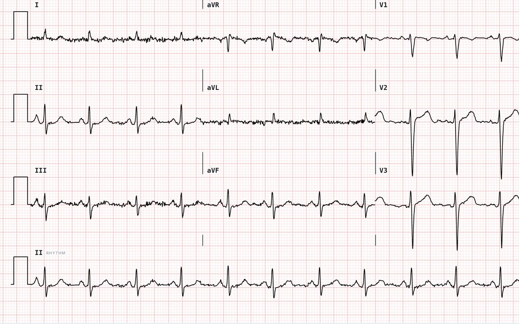
21,000+ real 12-leads
The full 12-lead library, every record measured, with calipers and a magnifier.
 EKG Quiz
Create account
A free ECG practice test with answers. Twenty questions that mix real rhythm strips with the EKG basics, and a source behind every answer.
Loading the first question
Your results
Review the ones you missed
Every quiz, the full flashcard decks, Telemetry Watch and more.
$9.99 for 3 months
One payment, plus tax where it applies. Never renews, nothing to cancel. Back to Free when it ends, with your progress kept.
Get the 3-month pass$29 a year
Billed annually, plus tax where it applies. Renews automatically each year. Cancel anytime.
Start ProEverything in Pro
Not ready yet? Create a free account, no card needed.
EKGLab is ECG training built on real patient recordings, with quizzes, flashcards and live tools. A free account needs no card.
The full 12-lead library, every record measured, with calipers and a magnifier.
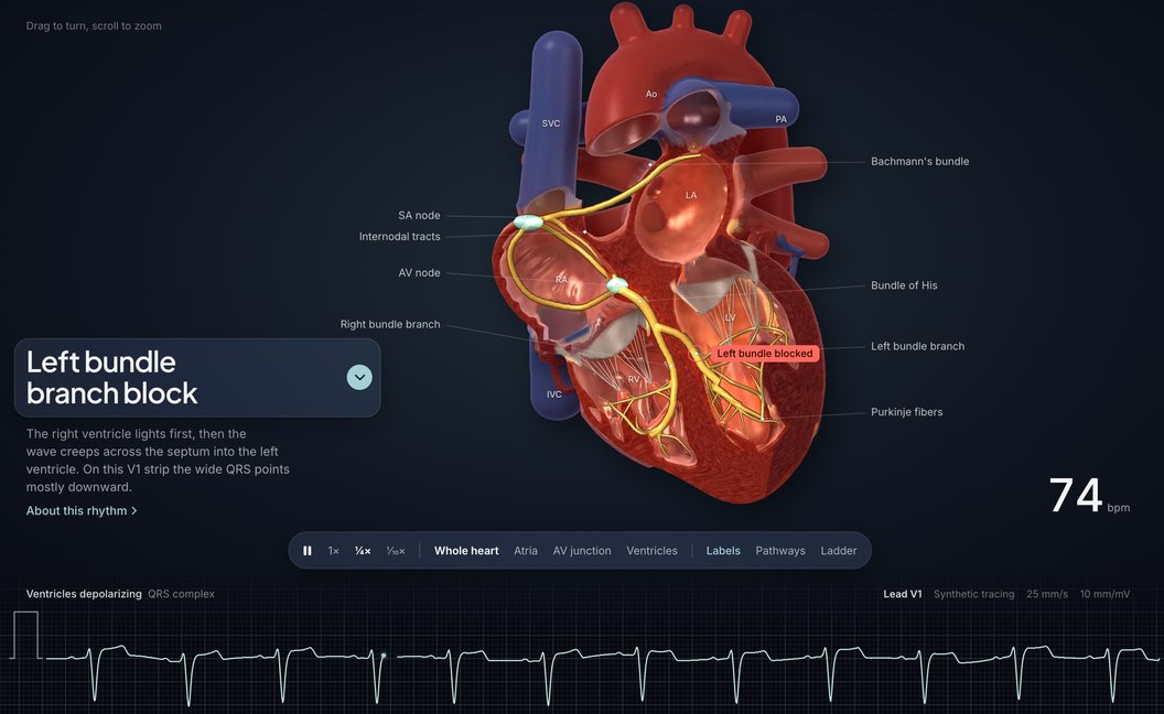
Watch 16 rhythms travel through a 3D heart while the strip writes itself.
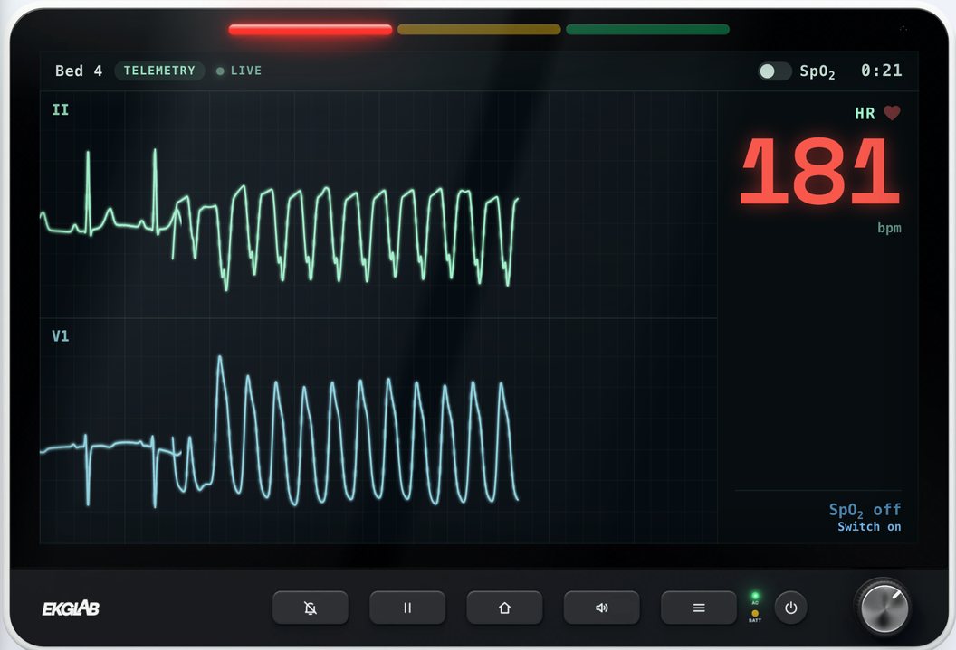
Catch the moment a rhythm changes on a live bedside monitor.

Rhythms, heart blocks, ectopy, ST changes, Q waves and bundle branch blocks.
This EKG quiz mixes reading real tracings with the fundamentals every ECG reader needs. Each run is 20 questions, 8 on rhythm strips and 12 on the EKG basics, in these areas:
Every strip is a real recording, shown at 25 mm/s and 10 mm/mV, with calipers, a magnifier and the whole 12-lead one click away. These are the ECG rhythms you may meet, each linked to its definition in the EKG Dictionary.
EKG Practice Strips has more real rhythm strips to read step by step, with answers and no sign-up. The EKG Cheat Sheet puts sixteen rhythms on one printable page. Rhythm Foundations teaches eleven common ECG rhythms, and the EKG Dictionary defines each term with its sources.
Yes. They are two names for the same test, the electrocardiogram. EKG comes from the German spelling and is common in the United States; ECG is used in most other countries. EKGLab uses both.
Yes. The quiz, its answers and its sources are free, and you do not need an account. Nothing you answer is saved or sent.
Each run is 20 questions: 8 on real ECG strips and 12 on the EKG basics, drawn at random, so you can take it more than once. Every EKG basics question in the bank is listed on this page with its answer.
Yes. Every strip has calipers for measuring intervals and a magnifier, and a button that opens the whole 12-lead ECG the strip came from.
EKGLab's quizzes pass at 80%.
Every EKG basics answer shows its sources: hover over or tap a small number in an explanation to read the source's own words. Every strip is a real recording, checked against the measurements the 12-lead machine made on it, with the EKG Dictionary's definition and sources after you answer.
EKGLab is practice: reading real rhythm strips and 12-leads, with feedback on every answer. It is not a certification course and does not award a certificate. This quiz covers the fundamentals. It is not a certification exam such as the NHA CET, and EKGLab is not affiliated with any certifying body.
Every EKG basics question in the bank (40), with its answer, its explanation and its sources.
Answer: 25 millimeters per second
The usual standard paper speed is 25 millimeters per second. Some countries use 50 millimeters per second as standard, and the rule for working out the heart rate changes with it.
Sources: Meek 2002a, BMJ; Sattar 2023, StatPearls
Answer: One small box is 0.04 seconds wide, and one large box is 0.20 seconds wide.
At 25 millimeters per second a small box (1 millimeter) lasts 0.04 seconds. A large box is five small boxes wide, so it lasts 0.20 seconds.
Sources: Meek 2002a, BMJ
Answer: 5 large boxes
Each large box lasts 0.20 seconds, so five large boxes make one full second.
Sources: Meek 2002a, BMJ
Answer: 10 millimeters (two large boxes)
The standard calibration is 10 millimeters per millivolt, so a 1 millivolt signal stands 10 millimeters tall, which is two large boxes. Analog machines need the calibration verified, and computerized machines verify it automatically.
Answer: True
At 10 millimeters per millivolt, each small box is 0.1 millivolt tall, so the five small boxes of one large box add up to 0.5 millivolt.
Sources: Meek 2002a, BMJ
Answer: Left to right is time, and up and down is voltage
Time runs from left to right, so the width of a wave is how long it lasts, in seconds. Height is voltage (also called amplitude), measured in millivolts.
Sources: Sattar 2023, StatPearls; Meek 2002a, BMJ
Answer: 10 seconds
A standard 12-lead printout covers 10 seconds. In the usual layout each of its four columns shows 2.5 seconds of three leads, and a rhythm strip along the bottom, when the page has one, runs the full 10 seconds.
Sources: Sattar 2023, StatPearls; Kligfield 2007, Circulation
Match each with its answer:
Answer: P wave: Atrial depolarization; QRS complex: Ventricular depolarization; T wave: Ventricular repolarization
The P wave is atrial depolarization, the QRS complex is ventricular depolarization and the T wave is ventricular repolarization.
Sources: Meek 2002b, BMJ
Match each with its answer:
Answer: Q wave: The first downward deflection, before any R wave; R wave: An upward deflection; S wave: A downward deflection that follows an R wave
The waves of a QRS complex are named by their direction and their order: a Q wave is a downward deflection that comes before any R wave, an R wave is an upward deflection, and an S wave is a downward deflection that follows an R wave. Not every QRS complex has all three: the S wave, for example, is often absent in leads V5 and V6.
Sources: Meek 2002b, BMJ
Answer: Depolarization
Depolarization is the electrical activation of the heart muscle, and it is what sets off contraction. Repolarization is its electrical recovery, which leaves the cells ready for the next beat.
Sources: Meek 2002a, BMJ; Wei 2023, StatPearls; Open RN 2023, Nursing Advanced Skills; Rogoff 2022, StatPearls
Answer: U wave
The U wave is a small, low deflection that may follow the T wave. Many EKGs show no clear U wave, and when it is present it is seen best in leads V2 and V3 and at slower heart rates.
Sources: Rautaharju 2009, Circulation; Meek 2002b, BMJ
Answer: J point
The J point is the junction where the QRS complex ends and the ST segment begins. ST elevation and ST depression are usually measured there, against the baseline.
Sources: Meek 2002b, BMJ; Rautaharju 2009, Circulation
Answer: From the start of the P wave to the start of the QRS complex
The PR interval runs from the start of the P wave to the start of the QRS complex, whether the QRS complex begins with a Q wave or an R wave. The flat stretch from the end of the P wave to the QRS complex is the PR segment.
Sources: Meek 2002b, BMJ
Answer: The PR segment is the stretch of line between the P wave and the QRS complex. The PR interval runs from the start of the P wave, so it includes the P wave.
The PR segment is the line after the P wave, up to the QRS complex. The PR interval starts at the beginning of the P wave, so it contains that wave. The ST segment and the QT interval differ in the same way: the ST segment is the line between the QRS complex and the T wave, and the QT interval includes both of those waves.
Sources: Meek 2002b, BMJ
Answer: From the end of the QRS complex to the start of the T wave
The ST segment runs from the end of the QRS complex to the start of the T wave. It is normally about level with the baseline, though some elevation at its start is normal in most chest leads.
Sources: Meek 2002b, BMJ; Wagner 2009, Circulation; Rautaharju 2009, Circulation
Answer: From the start of the QRS complex to the end of the T wave
The QT interval runs from the start of the QRS complex to the end of the T wave. It covers the whole of ventricular depolarization and repolarization.
Sources: Meek 2002b, BMJ
Answer: Because the QT interval changes with the heart rate
The QT interval lengthens as the heart rate slows and shortens as it speeds up. A formula such as Bazett's corrects it for the rate, and the result is written QTc.
Sources: Meek 2002b, BMJ; Sattar 2023, StatPearls
Answer: The time between two consecutive R waves
The R-R interval is the time between two consecutive R waves. It is the basis for working out the ventricular rate.
Sources: Meek 2002b, BMJ; Meek 2002a, BMJ
Answer: The flat baseline of the tracing, against which the waves are measured
The isoelectric line is the flat baseline of the tracing. Waves are read as upward or downward from it, and ST segment shifts and T wave heights are measured against the TP or PR segment, which normally lie on it.
Sources: Open RN 2023, Nursing Advanced Skills; Rautaharju 2009, Circulation; Meek 2002a, BMJ; Meek 2002b, BMJ
Answer: False
The atrial repolarization wave, called the Ta wave, is usually hidden by the much larger QRS complex and T wave. So a normal EKG does not show it as a separate wave. It can be recorded in third-degree AV block, when the Ta wave and the QRS complexes are uncoupled.
Sources: Holmqvist 2009, Ann Noninvasive Electrocardiol; Wei 2023, StatPearls
Answer: 0.12 to 0.20 seconds
The normal adult PR interval is 0.12 to 0.20 seconds (120 to 200 milliseconds), which is three to five small boxes at the standard paper speed, and it varies with age and heart rate.
Answer: 0.16 seconds
Each small box is 0.04 seconds, so four small boxes are 0.16 seconds (160 milliseconds).
Sources: Meek 2002a, BMJ
Answer: 0.12 seconds (three small boxes)
A QRS complex that lasts 0.12 seconds (120 milliseconds, three small boxes) or more is called wide. A QRS complex shorter than that is usually called narrow.
Sources: Meek 2002b, BMJ; Samesima 2022, Arq Bras Cardiol; Sattar 2023, StatPearls
Answer: A heart rate below 60 beats per minute is called bradycardia, and a heart rate above 100 beats per minute is called tachycardia.
By the traditional definitions, bradycardia is a heart rate below 60 beats per minute and tachycardia is a heart rate above 100 beats per minute. For its own purposes the 2018 bradycardia guideline uses a lower figure, a sinus rate below 50 beats per minute, and the 2022 Brazilian guideline defines sinus bradycardia the same way.
Sources: Meek 2002a, BMJ; Kusumoto 2019, Circulation; Samesima 2022, Arq Bras Cardiol
Answer: 75 beats per minute
Divide 300 by the number of large boxes between two consecutive R waves: 300 divided by 4 is 75 beats per minute.
Sources: Meek 2002a, BMJ
Answer: P waves
The atrial rate is the number of P waves per minute, and the ventricular rate is the number of QRS complexes per minute. In a normal rhythm a P wave comes before each QRS complex, so the two are the same, but a dysrhythmia can make them differ.
Sources: Open RN 2023, Nursing Advanced Skills; Rogoff 2022, StatPearls
Answer: 70 beats per minute
For an irregular rhythm such as atrial fibrillation, count the QRS complexes in a 6 second strip (30 large boxes) and multiply by 10: 7 times 10 is 70 beats per minute.
Sources: Open RN 2023, Nursing Advanced Skills; Samesima 2022, Arq Bras Cardiol
Answer: False
The 12 leads come from 10 electrodes: 4 on the limbs and 6 on the chest.
Sources: Kligfield 2007, Circulation; Sattar 2023, StatPearls
Sort into Limb leads and Chest (precordial) leads:
Answer: Lead I: Limb leads; V6: Chest (precordial) leads; Lead III: Limb leads; V3: Chest (precordial) leads; aVL: Limb leads; V1: Chest (precordial) leads
The six limb leads are I, II, III, aVR, aVL and aVF, and they view the heart in the frontal (vertical) plane. The six chest (precordial) leads are V1 through V6, and they view it in the horizontal plane.
Sources: Meek 2002a, BMJ; Kligfield 2007, Circulation
Answer: An upward (positive) deflection
Depolarization that travels toward the positive electrode of a lead is recorded as an upward deflection, and depolarization that travels away from it as a downward deflection.
Sources: Kligfield 2007, Circulation; Sattar 2023, StatPearls
Answer: Leads II, III and aVF
Leads II, III and aVF view the inferior surface of the heart. Leads I, aVL, V5 and V6 view the lateral surface, and leads V1 through V4 view the anterior surface. Some references call V1 and V2 the septal leads, and V3 and V4 the anterior leads.
Sources: Meek 2002a, BMJ; Open RN 2023, Nursing Advanced Skills
Answer: In the fourth intercostal space at the right border of the sternum
V1 is placed in the fourth intercostal space at the right border of the sternum, and V2 in the same space at the left border. V4 is placed in the fifth intercostal space in the midclavicular line.
Sources: Kligfield 2007, Circulation
Answer: II
Lead II is the usual choice for a rhythm strip, because it usually gives a good view of the P wave.
Sources: Meek 2002a, BMJ
Answer: True
Misplaced chest electrodes can change the waves on a tracing a great deal. Placing V1 and V2 too high can shrink the first R wave in those leads. A related error, swapping the left and right arm wires, inverts lead I. Limb lead switches can also give false-positive and false-negative signs of ischemia.
Sources: Kligfield 2007, Circulation
Answer: False
Not every printed EKG is diagnostic quality: filtering to a band of 1 to 30 Hz gives a stable tracing, but that bandwidth is unacceptable for diagnostic recording. With traditional analog filters, a 0.5 Hz low-frequency cutoff, once widely used in rhythm monitors, can distort repolarization and produce false ST deviation. To reduce that distortion, the AHA standard sets the low-frequency cutoff at 0.05 Hz, or at 0.67 Hz or below for digital filters designed to avoid it.
Sources: Kligfield 2007, Circulation
Answer: 1. Sinoatrial (SA) node; 2. Atrioventricular (AV) node; 3. Bundle of His; 4. Right and left bundle branches; 5. Purkinje fibers
A normal impulse begins in the sinoatrial node and spreads across the atria to the atrioventricular node, where it pauses briefly. It then travels down the bundle of His, through the right and left bundle branches and into the Purkinje fibers, which activate the ventricles.
Sources: Meek 2002a, BMJ; Rogoff 2022, StatPearls
Match each with its answer:
Answer: Sinoatrial (SA) node: 60 to 100 beats per minute; Atrioventricular (AV) junction: 40 to 60 beats per minute; Ventricles: 20 to 40 beats per minute
The sinoatrial node fires fastest, at about 60 to 100 beats per minute, so it normally sets the pace. If its impulses fail to form or to arrive, a slower site lower down can take over: about 40 to 60 beats per minute from the atrioventricular junction and about 20 to 40 from the ventricles. Sources differ a little on the lower ranges: one gives 40 to 50 for the junction and 30 to 40 for the ventricles.
Sources: Wei 2023, StatPearls; Sattar 2023, StatPearls; Open RN 2023, Nursing Advanced Skills; Rogoff 2022, StatPearls; Ashkanani 2026, Ann Noninvasive Electrocardiol
Answer: A P wave before every QRS complex, A rate of 60 to 100 beats per minute, Upright P waves in lead II
Normal sinus rhythm begins in the sinoatrial node. It has upright P waves in leads I and II, a P wave before every QRS complex and a PR interval of 0.12 to 0.20 seconds. By the traditional definition its rate is 60 to 100 beats per minute, and it is regular apart from the normal change with breathing.
Sources: Meek 2002a, BMJ; Rogoff 2022, StatPearls; Meek 2002b, BMJ; Samesima 2022, Arq Bras Cardiol
Answer: The ability of certain heart cells to start an impulse on their own
Automaticity is the ability of specialized heart cells to depolarize spontaneously and start an impulse without any outside stimulus. The sinoatrial node normally has the highest built-in rate, so it leads.
Sources: Wei 2023, StatPearls; Rogoff 2022, StatPearls; Sattar 2023, StatPearls
Answer: Deflections on the tracing that do not come from the heart’s electrical activity
Artifact is anything on the tracing that does not come from the heart’s electrical activity. Common sources are movement, muscle tremor, electrical interference and loose electrodes.
Sources: Pérez-Riera 2018, Ann Noninvasive Electrocardiol; Open RN 2023, Nursing Advanced Skills; Kligfield 2007, Circulation